
ଫାଇଲ:Pulmonary contusion CT arrow.jpg
ଏହି ଦେଖଣାର ଆକାର: ୮୦୦ × ୫୮୮ ପିକ୍ସେଲ । ବାକି ରେଜୋଲୁସନ: ୩୨୦ × ୨୩୫ ପିକ୍ସେଲ | ୬୪୦ × ୪୭୦ ପିକ୍ସେଲ | ୮୫୩ × ୬୨୭ ପିକ୍ସେଲ.
ମୂଳ ଫାଇଲ (୮୫୩ × ୬୨୭ ପିକସେଲ, ଫାଇଲ ଆକାର: ୯୪ KB, ଏମ.ଆଇ.ଏମ.ଇର ପ୍ରକାର: image/jpeg)
This is a file from the Wikimedia Commons. Information from its description page there is shown below. |
| ବିବରଣୀ |
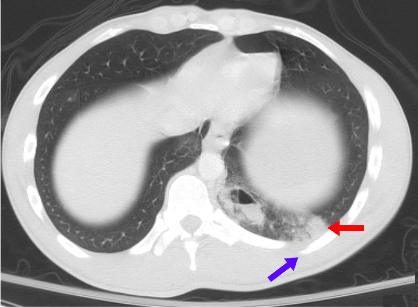
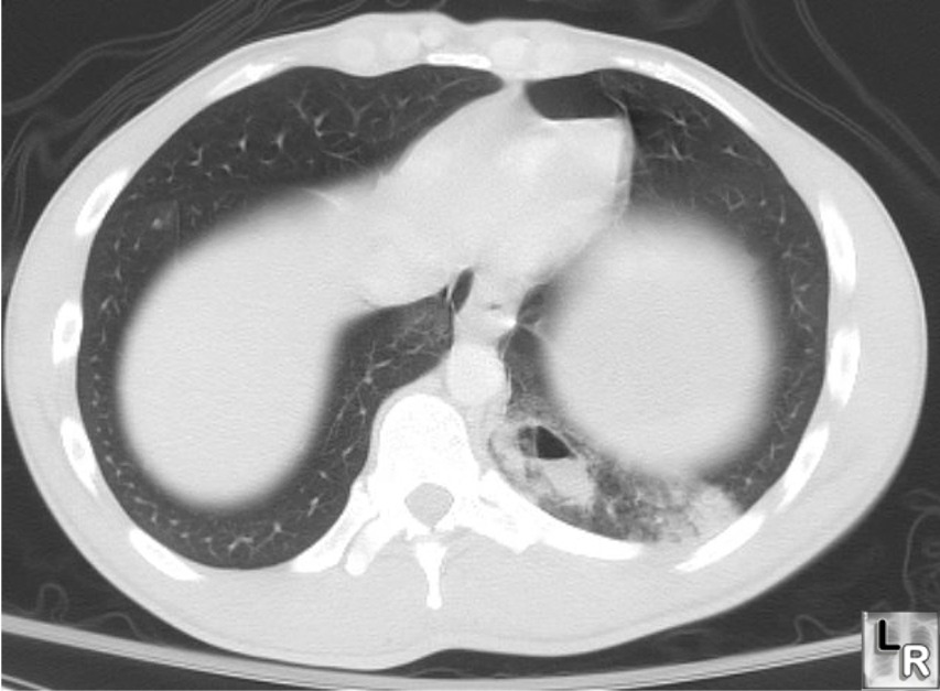
English: Modified version of Image:Pulmonary contusion CT.jpg, a CT scan of a patient with a "partially-cystic, partially fluid-filled structure" (dark spot near spine on middle-right hand lower portion of image), a rib fracture (lower righthand portion of image), and "subpleural hemorrhage representing a pulmonary contusion (lighter area near rib fracture in lower right hand portion of image, indicated by red arrow), as described at LearningRadiology.com. Blue arrow points to rib fracture. |
| ତାରିଖ | modified 6/23/08 |
| ମୂଳାଧାର | from LearningRadiology.com. Original context. Modified by version of Image:Pulmonary contusion CT.jpg |
| ଲେଖକ | LearningRadiology.com |
| ଅନୁମତି (ଏହି ଫାଇଲକୁ ପୁଣି ବ୍ୟବହାର କରିବେ) |
cc-by-sa, attribute to LearningRadiology.com. Modified from Image:Pulmonary contusion CT.jpg, for which the ticket number is copied below |
| ଅନ୍ୟ ସଂସ୍କରଣ | Image:Pulmonary contusion CT.jpg |
ଏହି ଫାଇଲଟି କ୍ରିଏଟିଭ କମନ୍ସ ଅଧୀନରେ ଆଟ୍ରିବୁସନ ସେଆର-ଏଲାଇକ ୩.୦ ଅନପୋର୍ଟେଡ଼ ଲାଇସେନ୍ସରେ ପଞ୍ଜିକରଣ କରାଯାଇଅଛି ।
ଶ୍ରେୟ: Reusers must attribute LearningRadiology.com for the original image
- ଆପଣ ଆରାମରେ:
- ବାଣ୍ଟିପାରିବେ – କାମଟିକୁ ନକଲ କରିପାରିବେ, ବାଣ୍ଟିପାରିବେ ଓ ପ୍ରସାରଣ କରିପାରିବେ
- ମିଶାଇପାରିବେ – କାମଟି ଅଭିଯୋଜନ କରିପାରିବେ
- ତଳଲିଖିତ ସର୍ତ୍ତାବଳୀ ଅଧୀନରେ:
- ଶ୍ରେୟ – ଆପଣ ମନେ କରି ଏହି କାମର ଆବଶ୍ୟକୀୟ ଶ୍ରେୟ ମୂଳ ଗଢ଼ାଳି ବା ସ୍ୱତ୍ୱାଧୀକାରୀଙ୍କୁ ଦେବେ ଏବଂ ଦେଲାବେଳେ ଲାଇସେନ୍ସର ଲିଙ୍କ ଦେଇ କି କି ବଦଳ କଲେ ଉଲ୍ଲେଖ କରିବେ । ଏହା ଉପଯୁକ୍ତ ଢଙ୍ଗରେ କରିବେ କିନ୍ତୁ ଲାଇସେନ୍ସ ଦେଉଥିବା ବ୍ୟକ୍ତି ଆପଣଙ୍କ ପ୍ରଚାର କଲା ଭଳି କିଛି ଲେଖିବେ ନାହିଁ ।
- ସେଆର ଏଲାଇକ – ଯଦି ଆପଣ ଏହି କାମଟିକୁ ବଦଳାଇବେ, ରୂପାନ୍ତରଣ କରିବେ ବା ଏହାକୁ ନେଇ କିଛି ଗଢ଼ିବେ ତେବେ ଆପଣ ଏହାକୁ ଏକା ବା ଅଲଗା ଲାଇସେନ୍ସ ଭିତରେ ରଖିପାରିବେ ।
ଫାଇଲ ଇତିହାସ
ଏହା ଫାଇଲଟି ସେତେବେଳେ ଯେମିତି ଦିଶୁଥିଲା ତାହା ଦେଖିବା ପାଇଁ ତାରିଖ/ବେଳା ଉପରେ କ୍ଲିକ କରନ୍ତୁ
| ତାରିଖ/ବେଳ | ନଖ ଦେଖଣା | ଆକାର | ବ୍ୟବହାରକାରୀ | ମତାମତ | |
|---|---|---|---|---|---|
| ଏବେକାର | ୦୫:୦୬, ୨୪ ଜୁନ ୨୦୦୮ | ୮୫୩ × ୬୨୭ (୯୪ KB) | Delldot | {{Information |Description={{en|1=Modified version of Image:Pulmonary contusion CT.jpg, a CT scan of a patient with a "partially-cystic, partially fluid-filled structure" (dark spot near spine on middle-right hand lower portion of image), a [[rib fra |
ଫାଇଲ ବ୍ୟବହାର
ଏହି ସବୁପୃଷ୍ଠା ଏହି ଫାଇଲଟିକୁ ଯୋଡ଼ିଥାନ୍ତି:
ଜଗତ ଫାଇଲ ବ୍ୟବହାର
ତଳଲିଖିତ ଉଇକିସବୁ ଏହି ଫାଇଲଟିକୁ ବ୍ୟବହାର କରିଥାନ୍ତି:
- ar.wikipedia.orgରେ ବ୍ୟବହାର
- ast.wikipedia.orgରେ ବ୍ୟବହାର
- bn.wikipedia.orgରେ ବ୍ୟବହାର
- ca.wikipedia.orgରେ ବ୍ୟବହାର
- en.wikipedia.orgରେ ବ୍ୟବହାର
- es.wikipedia.orgରେ ବ୍ୟବହାର
- fa.wikipedia.orgରେ ବ୍ୟବହାର
- fr.wikipedia.orgରେ ବ୍ୟବହାର
- he.wikipedia.orgରେ ବ୍ୟବହାର
- id.wikipedia.orgରେ ବ୍ୟବହାର
- it.wikipedia.orgରେ ବ୍ୟବହାର
- ja.wikipedia.orgରେ ବ୍ୟବହାର
- ko.wikipedia.orgରେ ବ୍ୟବହାର
- nn.wikipedia.orgରେ ବ୍ୟବହାର
- pt.wikipedia.orgରେ ବ୍ୟବହାର
- sr.wikipedia.orgରେ ବ୍ୟବହାର
- th.wikipedia.orgରେ ବ୍ୟବହାର
- tr.wikipedia.orgରେ ବ୍ୟବହାର
- www.wikidata.orgରେ ବ୍ୟବହାର
- zh.wikipedia.orgରେ ବ୍ୟବହାର