
ଫାଇଲ:Fundus of patient with retinitis pigmentosa, mid stage.jpg
ଏହି ଦେଖଣାର ଆକାର: ୬୯୯ × ୫୯୯ ପିକ୍ସେଲ । ବାକି ରେଜୋଲୁସନ: ୨୮୦ × ୨୪୦ ପିକ୍ସେଲ | ୫୬୦ × ୪୮୦ ପିକ୍ସେଲ | ୮୭୧ × ୭୪୭ ପିକ୍ସେଲ.
ମୂଳ ଫାଇଲ (୮୭୧ × ୭୪୭ ପିକସେଲ, ଫାଇଲ ଆକାର: ୧୦୭ KB, ଏମ.ଆଇ.ଏମ.ଇର ପ୍ରକାର: image/jpeg)
This is a file from the Wikimedia Commons. Information from its description page there is shown below. |
| ବିବରଣୀ |
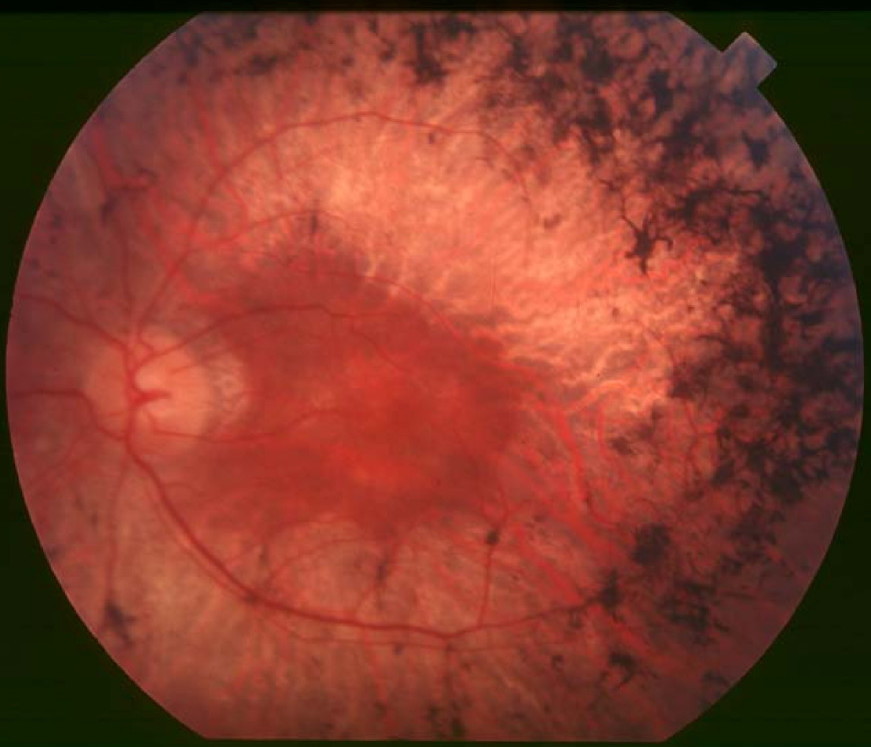
English: Figure 2. Fundus of patient with retinitis pigmentosa, mid stage (Bone spicule-shaped pigment deposits are present in the mid periphery along with retinal atrophy, while the macula is preserved although with a peripheral ring of depigmentation. Retinal vessels are attenuated.) Hamel Orphanet Journal of Rare Diseases 2006 1:40 doi:10.1186/1750-1172-1-40 |
| ତାରିଖ | |
| ମୂଳାଧାର | Retinitis pigmentosa by Christian Hamel |
| ଲେଖକ | Christian Hamel |
| ଅନୁମତି (ଏହି ଫାଇଲକୁ ପୁଣି ବ୍ୟବହାର କରିବେ) |
© 2006 Hamel; licensee BioMed Central Ltd. This is an Open Access article distributed under the terms of the Creative Commons Attribution License (https://creativecommons.org/licenses/by/2.0), which permits unrestricted use, distribution, and reproduction in any medium, provided the original work is properly cited. |
ଏହି ଫାଇଲଟି କ୍ରିଏଟିଭ କମନ୍ସ ଅଧୀନରେ ଆଟ୍ରିବୁସନ ୨.୦ ଜେନେରିକ ଲାଇସେନ୍ସରେ ପଞ୍ଜିକରଣ କରାଯାଇଅଛି ।
- ଆପଣ ଆରାମରେ:
- ବାଣ୍ଟିପାରିବେ – କାମଟିକୁ ନକଲ କରିପାରିବେ, ବାଣ୍ଟିପାରିବେ ଓ ପ୍ରସାରଣ କରିପାରିବେ
- ମିଶାଇପାରିବେ – କାମଟି ଅଭିଯୋଜନ କରିପାରିବେ
- ତଳଲିଖିତ ସର୍ତ୍ତାବଳୀ ଅଧୀନରେ:
- ଶ୍ରେୟ – ଆପଣ ମନେ କରି ଏହି କାମର ଆବଶ୍ୟକୀୟ ଶ୍ରେୟ ମୂଳ ଗଢ଼ାଳି ବା ସ୍ୱତ୍ୱାଧୀକାରୀଙ୍କୁ ଦେବେ ଏବଂ ଦେଲାବେଳେ ଲାଇସେନ୍ସର ଲିଙ୍କ ଦେଇ କି କି ବଦଳ କଲେ ଉଲ୍ଲେଖ କରିବେ । ଏହା ଉପଯୁକ୍ତ ଢଙ୍ଗରେ କରିବେ କିନ୍ତୁ ଲାଇସେନ୍ସ ଦେଉଥିବା ବ୍ୟକ୍ତି ଆପଣଙ୍କ ପ୍ରଚାର କଲା ଭଳି କିଛି ଲେଖିବେ ନାହିଁ ।
ଫାଇଲ ଇତିହାସ
ଏହା ଫାଇଲଟି ସେତେବେଳେ ଯେମିତି ଦିଶୁଥିଲା ତାହା ଦେଖିବା ପାଇଁ ତାରିଖ/ବେଳା ଉପରେ କ୍ଲିକ କରନ୍ତୁ
| ତାରିଖ/ବେଳ | ନଖ ଦେଖଣା | ଆକାର | ବ୍ୟବହାରକାରୀ | ମତାମତ | |
|---|---|---|---|---|---|
| ଏବେକାର | ୧୫:୪୭, ୨ ଡିସେମ୍ବର ୨୦୧୭ | | ୮୭୧ × ୭୪୭ (୧୦୭ KB) | Doc James | Cropped 27 % horizontally and 7 % vertically using CropTool with precise mode. |
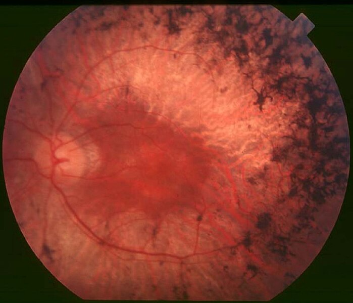
| ୧୯:୨୨, ୨୨ ସେପ୍ଟେମ୍ବର ୨୦୦୯ |  | ୧,୨୦୦ × ୭୯୯ (୧୨୬ KB) | CopperKettle | {{Information |Description={{en|1=Figure 2. Fundus of patient with retinitis pigmentosa, mid stage (Bone spicule-shaped pigment deposits are present in the mid periphery along with retinal atrophy, while the macula is preserved although with a peripheral |
ଫାଇଲ ବ୍ୟବହାର
ଏହି ସବୁପୃଷ୍ଠା ଏହି ଫାଇଲଟିକୁ ଯୋଡ଼ିଥାନ୍ତି:
ଜଗତ ଫାଇଲ ବ୍ୟବହାର
ତଳଲିଖିତ ଉଇକିସବୁ ଏହି ଫାଇଲଟିକୁ ବ୍ୟବହାର କରିଥାନ୍ତି:
- ar.wikipedia.orgରେ ବ୍ୟବହାର
- bs.wikipedia.orgରେ ବ୍ୟବହାର
- ca.wikipedia.orgରେ ବ୍ୟବହାର
- da.wikipedia.orgରେ ବ୍ୟବହାର
- en.wikipedia.orgରେ ବ୍ୟବହାର
- en.wikiversity.orgରେ ବ୍ୟବହାର
- es.wikipedia.orgରେ ବ୍ୟବହାର
- eu.wikipedia.orgରେ ବ୍ୟବହାର
- fa.wikipedia.orgରେ ବ୍ୟବହାର
- fi.wikipedia.orgରେ ବ୍ୟବହାର
- fr.wikipedia.orgରେ ବ୍ୟବହାର
- he.wikipedia.orgରେ ବ୍ୟବହାର
- hy.wikipedia.orgରେ ବ୍ୟବହାର
- it.wikipedia.orgରେ ବ୍ୟବହାର
- ko.wikipedia.orgରେ ବ୍ୟବହାର
- la.wikipedia.orgରେ ବ୍ୟବହାର
- outreach.wikimedia.orgରେ ବ୍ୟବହାର
- pl.wikipedia.orgରେ ବ୍ୟବହାର
- pt.wikipedia.orgରେ ବ୍ୟବହାର
- ru.wikipedia.orgରେ ବ୍ୟବହାର
- sl.wikipedia.orgରେ ବ୍ୟବହାର
- sr.wikipedia.orgରେ ବ୍ୟବହାର
- sv.wikipedia.orgରେ ବ୍ୟବହାର
- th.wikipedia.orgରେ ବ୍ୟବହାର
- tr.wikipedia.orgରେ ବ୍ୟବହାର
- tt.wikipedia.orgରେ ବ୍ୟବହାର
- uk.wikipedia.orgରେ ବ୍ୟବହାର
- vi.wikipedia.orgରେ ବ୍ୟବହାର
ଏହି ଫାଇଲଟିର ଅଧିକ ବିଶ୍ୱବ୍ୟାପୀ ବ୍ୟବହାର ଦେଖନ୍ତୁ ।